Note
Go to the end to download the full example code.
Multi-tissue FODs of a multi-shell brain scan#
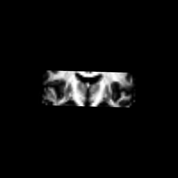
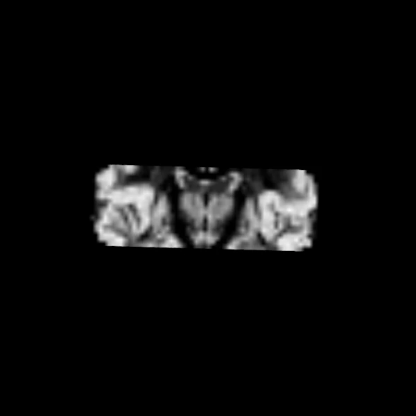
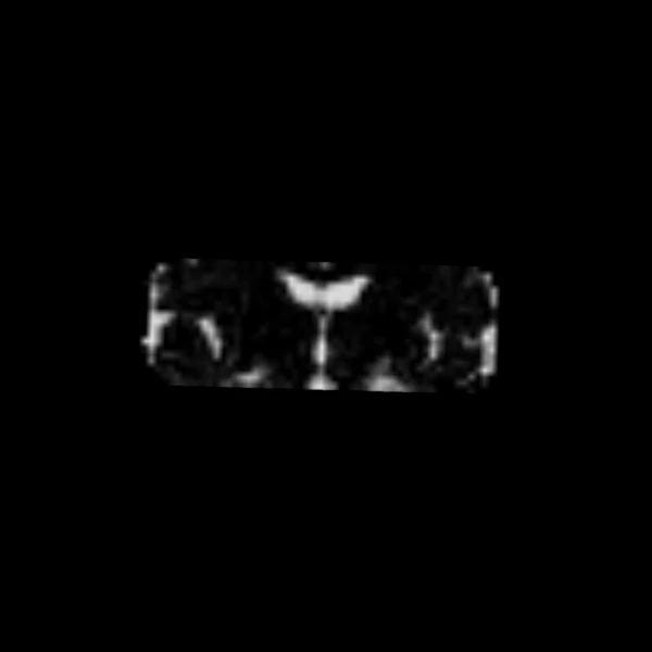
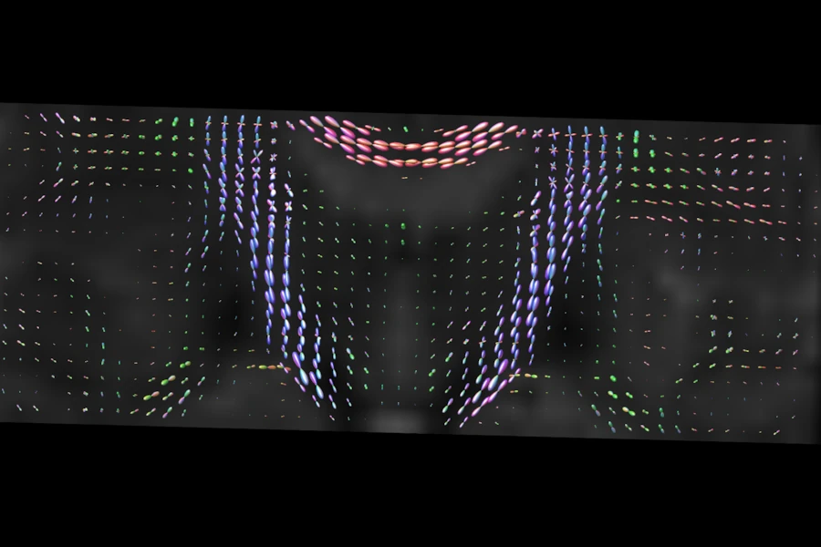
This example fits multi-shell multi-tissue constrained spherical deconvolution (MSMT-CSD) to a multi-shell scan of a healthy volunteer. The white matter, grey matter and CSF responses are estimated from the data itself, then each voxel gets a white matter FOD and the fraction of each tissue. It shows the FODs on a coronal slice and the three tissue fractions on the same slice.
The data is the CFIN multi-shell dataset (Hansen B, Jespersen SN. Data for evaluation of fast kurtosis strategies, b-value optimization and exploration of diffusion MRI contrast. Scientific Data 3 (2016) 160072).
MSMT-CSD follows Jeurissen B et al. NeuroImage 103 (2014) 411-426, and the tissue
responses are estimated as by dwi2response dhollander of MRtrix3 (Dhollander T et
al., 2016 and 2019; Tournier JD et al. NeuroImage 202 (2019) 116137).
# needs: example-data (the tests run it only when LARMOR_DATA_DIR is set)

import numpy as np

from larmor.data import fetch_cfin_multishell
from larmor.dwi import create_brain_mask, mean_b0
from larmor.io import load_dwi
from larmor.sd import MSMTCSDModel
from larmor.viewer import Category, Viewer, ViewKind
The data and the fit#
fetch_cfin_multishell downloads the data once and returns the paths of its
files. With no responses given, the model estimates them from the data in the
brain mask, then fits every voxel of the mask.
files = fetch_cfin_multishell()
dwi, gtab = load_dwi(files["dwi"], files["bvals"], files["bvecs"])
b0 = mean_b0(dwi, gtab)
mask = create_brain_mask(b0)
fit = MSMTCSDModel().fit(dwi, gtab, mask)
fod = dwi.with_data(fit.fods[0].astype(np.float32))
fractions = fit.tissue_fractions
assert fractions is not None
for name, i in (("white matter", 0), ("grey matter", 1), ("CSF", 2)):
    print(
        f"Mean {name} fraction in the brain: {np.nanmean(fractions[..., i][mask]):.2f}"
    )
Mean white matter fraction in the brain: 0.34
Mean grey matter fraction in the brain: 0.51
Mean CSF fraction in the brain: 0.15
White matter FODs#
The glyphs are drawn over the mean b=0 image, on a coronal slice through the centre of the brain.
shape = np.array(dwi.data.shape[:3])
centre = tuple(dwi.affine[:3, :3] @ ((shape - 1) / 2) + dwi.affine[:3, 3])

viewer = Viewer()
viewer.add(dwi.with_data(b0.astype(np.float32)), Category.VOLUMES, name="mean b=0")
viewer.add(fod, Category.FODS, name="WM FOD")
viewer.move_cursor(centre)
viewer.toggle_crosshair()
viewer.zoom(ViewKind.CORONAL, 2.0, about=centre)
viewer.screenshot(
    ViewKind.CORONAL, "msmt_csd_cfin_fods.png", size=(900, 600), overlay=False
)
Tissue fractions#
The fractions of white matter, grey matter and CSF in each voxel, on the same slice, each on its own.
for name, i, image in (
    ("WM fraction", 0, "msmt_csd_cfin_wm.png"),
    ("GM fraction", 1, "msmt_csd_cfin_gm.png"),
    ("CSF fraction", 2, "msmt_csd_cfin_csf.png"),
):
    viewer = Viewer()
    viewer.add(
        dwi.with_data(fractions[..., i].astype(np.float32)), Category.VOLUMES, name=name
    )
    viewer.move_cursor(centre)
    viewer.toggle_crosshair()
    viewer.screenshot(ViewKind.CORONAL, image, size=(600, 600), overlay=False)